
Flat moles, raised moles, atypical lesions you are uncertain about. Nexus Clinic KL assesses every mole before removal, selects the right method, and includes histopathology for any lesion with atypical features.
Every mole that bothers you deserves a proper medical assessment before anything else. The wrong removal method leaves a worse scar than the mole itself. At Nexus Clinic KL, our licensed aesthetic doctors assess every mole before recommending a removal method.
Precise Removal
CO2 Laser • RF • Surgical Excision
Nexus Clinic Kuala Lumpur — Excellence in Medical Aesthetics
Established
2001
Over 20 years of excellence
Location
Wisma UOA II, Jalan Pinang
KLCC, 50450 Kuala Lumpur
Opening Hours
Monday - Saturday
9:00am – 6:00pm | Closed Sundays & PH
MOH Registered & Compliant
All procedures performed by licensed doctors
Multiple Removal Methods
CO2 Laser • RF • Surgical Excision • Cryotherapy
Histopathology Available
For any lesion with atypical features
Everything you need to know
Methods Available
CO2 Laser Ablation, RF Cauterisation, Surgical Shave Excision, Surgical Excision with Sutures
Session Time
15 to 45 minutes depending on number and size of moles
Downtime
Minimal for laser and RF; 5 to 10 days healing for surgical excision
Sessions Required
Most moles cleared in 1 session; deeper moles may need 2 sessions
Anaesthesia
Local anaesthetic injection administered before all procedures
Histopathology
Tissue sample sent for lab analysis for any mole with atypical features
Suitable For
Flat moles, raised moles, compound moles, congenital moles; all skin types
Before any mole is removed, understand these clinical warning signs
One half does not match the other
Irregular, ragged, or blurred edges
Multiple shades of brown, tan, red, white, or blue
Larger than 6mm (size of a pencil eraser)
Changing in size, shape, colour, or symptoms
Important: Any mole with one or more ABCDE features requires clinical assessment before removal
At Nexus Clinic KL, moles with concerning features are excised with a margin of healthy surrounding tissue and sent for histopathological examination. This is not optional for any mole assessed as atypical.
Used at every initial mole assessment at Nexus Clinic KL
| Mole Type | Appearance | Depth | Histopathology Needed? | Recommended Method |
|---|---|---|---|---|
| Flat Junctional Naevus | Flat, evenly brown or black, well-defined border | Epidermis only | Not routinely required if no atypical features | CO2 Laser Ablation (1 session) |
| Raised Intradermal Naevus | Dome-shaped, flesh-coloured or lightly pigmented, soft | Dermis only | Not routinely required if benign appearance | RF Cauterisation or Surgical Shave Excision |
| Compound Naevus | Slightly raised, tan to brown, smooth surface | Epidermis and dermis | Recommended | Surgical Shave Excision or CO2 Laser; tissue sent for analysis |
| Congenital Naevus (Small) | Present from birth; tan to dark brown; may have hair | Variable | Recommended | Surgical Excision with Sutures; full specimen sent for histopathology |
| Dysplastic Naevus (Atypical Mole) | Irregular border, uneven colour, larger than 6mm | Variable; often deeper | Mandatory | Surgical Excision with Margins; histopathology compulsory |
| Seborrhoeic Keratosis (Warty Mole) | Rough, stuck-on appearance; waxy surface; light to dark brown | Epidermal layer | Not routinely required | CO2 Laser Ablation or RF Cauterisation (1 session) |
Mole Type
Flat Junctional Naevus
Appearance
Flat, evenly brown or black, well-defined border
Depth
Epidermis only
Histopathology Needed?
Not routinely required if no atypical features
Recommended Method
CO2 Laser Ablation (1 session)
Mole Type
Raised Intradermal Naevus
Mole Type
Compound Naevus
Mole Type
Congenital Naevus (Small)
Mole Type
Dysplastic Naevus (Atypical Mole)
Mole Type
Seborrhoeic Keratosis (Warty Mole)
Using surgical excision for a flat mole is the most common method mismatch in Malaysian mole removal. At Nexus Clinic KL, the correct method is selected based on clinical criteria rather than clinic preference.
What to expect during your recovery
Mild redness, tenderness, small wound. Local anaesthetic wears off. Keep area clean and dry.
Scab forms and begins to dry. No picking or scratching. Apply prescribed ointment.
Scab starts to separate naturally. New pink skin underneath. Continue sun protection.
Skin closes and calms. Mild pinkness may persist. Sunscreen essential daily.
Colour fades significantly. Scar continues to blend with surrounding skin.
Final result visible. Scar maturation complete. Continue sun protection.
Pro Tip: Sun protection over healed mole removal sites is important for at least 3 months after treatment. UV exposure on immature healing skin increases the risk of post-inflammatory pigmentation and slows scar fading.
What to expect based on your mole location and removal method
| Removal Method | Scarring Risk | Best Locations | Expected Healing Timeline |
|---|---|---|---|
| CO2 Laser Ablation | Very low; flat, skin-toned mark that fades over 4 to 8 weeks | Face, neck, chest; any visible area requiring minimal scarring | Pink mark visible for 2 to 4 weeks; full fading at 6 to 8 weeks |
| RF Cauterisation | Low; small flat scab forms and resolves; minimal permanent mark | Face, neck, back; suitable for raised benign moles | Scab falls off within 7 to 10 days; residual pinkness resolves at 4 to 6 weeks |
| Surgical Shave Excision | Low to moderate; flat scar forms at shave level; may retain mild colour | Raised moles on face, scalp and body; suitable for compound naevi | 7 to 14 days healing; scar maturation at 3 to 6 months |
| Surgical Excision with Sutures | Linear scar along incision line; fades significantly over 6 to 12 months | Deeper or atypical moles; body locations where appearance is less critical | Sutures removed at 7 to 14 days; full scar remodelling at 6 to 12 months |
Removal Method
CO2 Laser Ablation
Scarring Risk
Very low; flat, skin-toned mark that fades over 4 to 8 weeks
Best Locations
Face, neck, chest; any visible area requiring minimal scarring
Expected Healing Timeline
Pink mark visible for 2 to 4 weeks; full fading at 6 to 8 weeks
Removal Method
RF Cauterisation
Removal Method
Surgical Shave Excision
Removal Method
Surgical Excision with Sutures
Moles on the chest and upper back carry a higher risk of hypertrophic or keloid scarring in darker Fitzpatrick skin types. At Nexus Clinic KL, patients with a personal or family history of keloid scarring are counselled specifically about method choice.
Clinical accountability in mole removal
Removing a mole without histopathology when the clinical picture warrants it is a practice Nexus Clinic KL does not follow, regardless of the patient's stated preference. Patient safety is not optional.
Uses a precisely focused beam of carbon dioxide laser energy to vaporise mole tissue layer by layer. No cutting, no sutures, no incision. Treated area forms a flat protective crust that falls off within 7 to 10 days. Preferred method for flat junctional naevi and seborrhoeic keratoses on the face and visible body areas.
Uses a fine electrode through which controlled radiofrequency current passes, generating localised heat that precisely destroys mole tissue. Particularly well-suited for raised, dome-shaped intradermal naevi and skin tags. A small scab forms and separates naturally within 7 to 10 days.
Numbing the area with local anaesthetic and using a fine surgical blade to shave the raised portion of the mole flush with the surrounding skin. No sutures required. Used for compound naevi and situations where mole tissue needs to be preserved for histopathological examination.
Gold standard method for deep, large or atypical moles. Entire mole is cut out along with a margin of healthy surrounding tissue. Wound closed with fine sutures. Excised specimen sent for histopathological analysis in every case. The only technique that provides complete removal certainty.
Step by Step
Doctor examines every mole under bright lighting and dermoscopy. ABCDE criteria evaluated. Any atypical features documented and removal method selected with clear explanation.
Doctor explains recommended removal method, expected healing, realistic scar outcome, and whether histopathology will be performed. Written consent completed before procedure.
Local anaesthetic administered to each mole site. Takes effect within 2 to 3 minutes. Each mole takes 2 to 15 minutes to treat. Multiple moles treated in same session where appropriate.
Wound dressing applied. Written wound care instructions provided. Review appointment at 7 to 14 days for suture removal or wound assessment. Histopathology results reviewed at follow-up.
Slide to see the incredible transformations achieved by our clients
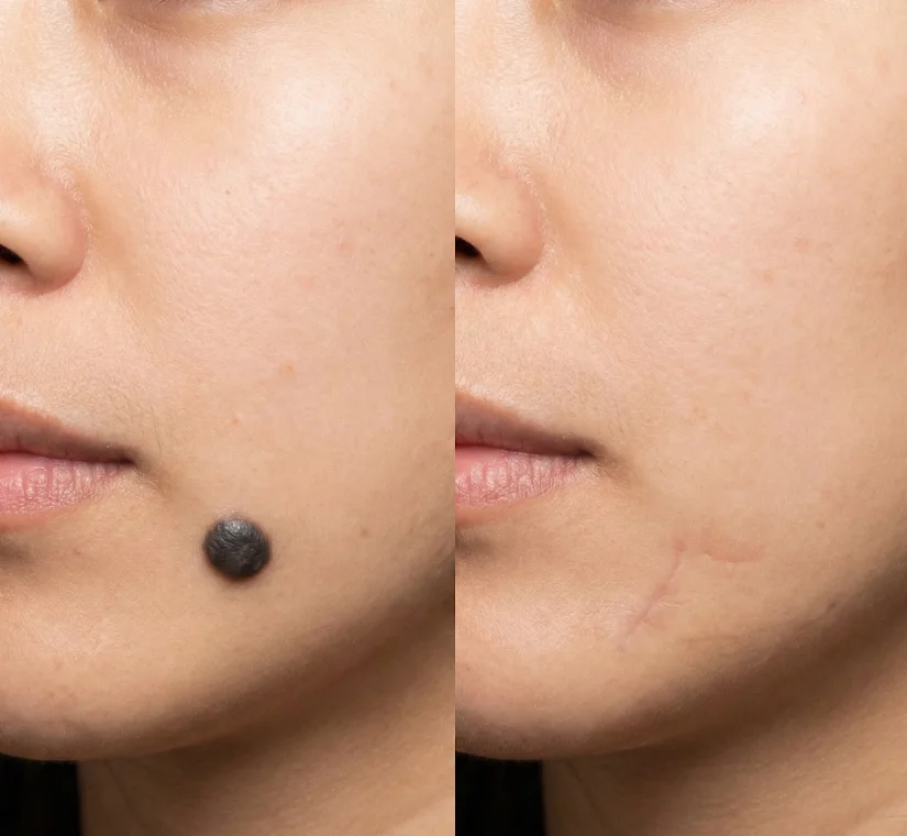
Drag to compare
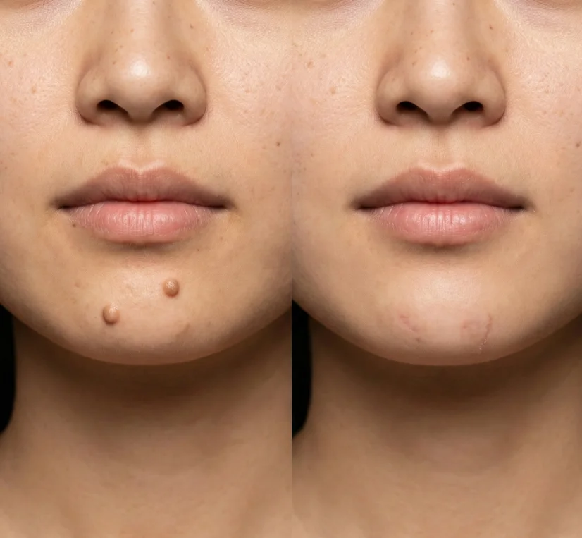
Drag to compare
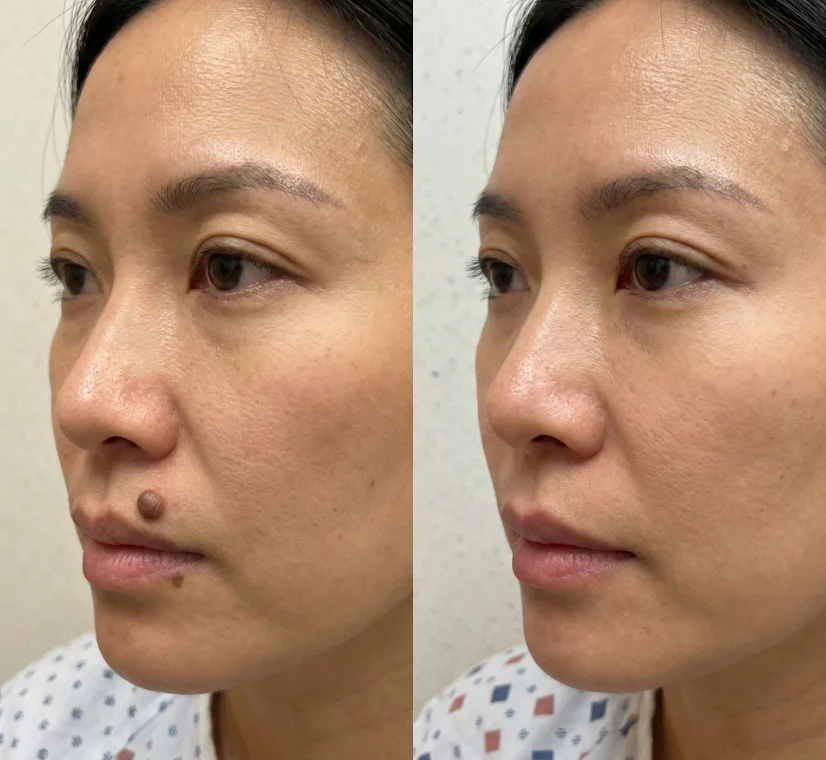
Drag to compare
Results may vary. Individual results depend on various factors.
Start Your TransformationTransparent pricing at Nexus Clinic KL
| Treatment / Method | Best Suited For | Sessions | Price Range (RM) 2026 |
|---|---|---|---|
| CO2 Laser Ablation (per mole) | Small to medium flat and raised benign moles | 1 (2 if deep) | RM 150 – RM 350 per mole |
| RF Cauterisation (per mole) | Raised benign moles and skin tags | 1 | RM 120 – RM 280 per mole |
| Surgical Shave Excision (per mole) | Raised compound naevi; moles requiring tissue sample | 1 | RM 250 – RM 500 per mole |
| Surgical Excision with Sutures (per mole) | Deep, large or atypical moles; histopathology indicated | 1 | RM 400 – RM 900 per mole (histopathology fee additional) |
| Multiple Moles Package (5 or more, CO2 or RF) | Multiple small benign moles in one session | 1 session | From RM 600 for 5 moles |
| Histopathology | Atypical, compound or suspicious moles | One-time with procedure | RM 150 – RM 350 (lab fee, separate from procedure) |
Treatment / Method
CO2 Laser Ablation (per mole)
Treatment / Method
RF Cauterisation (per mole)
Treatment / Method
Surgical Shave Excision (per mole)
Treatment / Method
Surgical Excision with Sutures (per mole)
Treatment / Method
Multiple Moles Package (5 or more, CO2 or RF)
Treatment / Method
Histopathology
Multiple mole removal in a single session offered at package pricing where clinically appropriate. Histopathology fees charged at cost and disclosed transparently before procedure.
Most mole removal is done with numbing medication, so the procedure itself should not be painful. You may feel mild stinging or burning for a few days afterward. The anaesthetic injection itself takes 2 to 3 seconds and involves a brief, sharp sensation. Once the area is numb, no pain is felt during the removal procedure.
Recovery is often minimal, with mild redness or scabbing for a few days to a week, depending on the method. Laser healing is commonly quoted around 7 to 14 days, while surgical excision may take longer. A small crust forms and falls off naturally within 7 to 10 days for laser and RF methods.
It can. Scar risk depends on mole depth and removal method. CO2 Laser Ablation leaves a flat, skin-toned mark that fades over 4 to 8 weeks. Surgical Excision with Sutures leaves a fine linear scar that fades over 6 to 12 months. Some people find the scar looks better than the mole once it fades.
Moles do not typically grow back unless they were incompletely removed. Moles removed by CO2 Laser Ablation or RF Cauterisation have a small possibility of partial recurrence. Moles removed by Surgical Excision with adequate margins virtually never recur.
Cost varies widely based on size, depth, location, and method. At Nexus Clinic KL, CO2 Laser Ablation starts from RM 150 per mole for small flat lesions. Surgical Excision with Sutures ranges from RM 400 to RM 900 per mole. Multiple mole packages starting from RM 600 for 5 moles are available.
When performed by a medical professional after proper assessment, laser removal can be a safe option for suitable moles. However, suspicious moles may need biopsy or a different pathway, so assessment comes first. CO2 Laser Ablation is particularly safe for flat, benign moles.
If it changes in size, shape, color, border, or starts evolving, get it checked. The ABCDE guide is a simple way to remember warning signs: Asymmetry, Border irregularity, Color variation, Diameter larger than 6mm, Evolution over time.
No. Professional medical organizations warn against at-home mole removal due to skin cancer risk, scarring, and infection. Never attempt to cut, burn, or use DIY removal creams on moles.
Not always. But if a mole looks suspicious, a doctor may take a tissue sample for microscopic examination. At Nexus Clinic KL, any mole with atypical features is sent for histopathological analysis.
There is no single 'best' method. It depends on depth, location, and your scar risk. CO2 Laser Ablation is often preferred for flat facial moles as it leaves minimal scarring. Surgical Shave Excision may be used for slightly raised moles.
Every mole is assessed before it is removed. The correct method is selected for the specific lesion type. Scar outcomes are discussed honestly based on your skin type and mole location.
Limited slots available this week | Located at Wisma UOA II, Jalan Pinang, KLCC — Serving Malaysia since 2001
